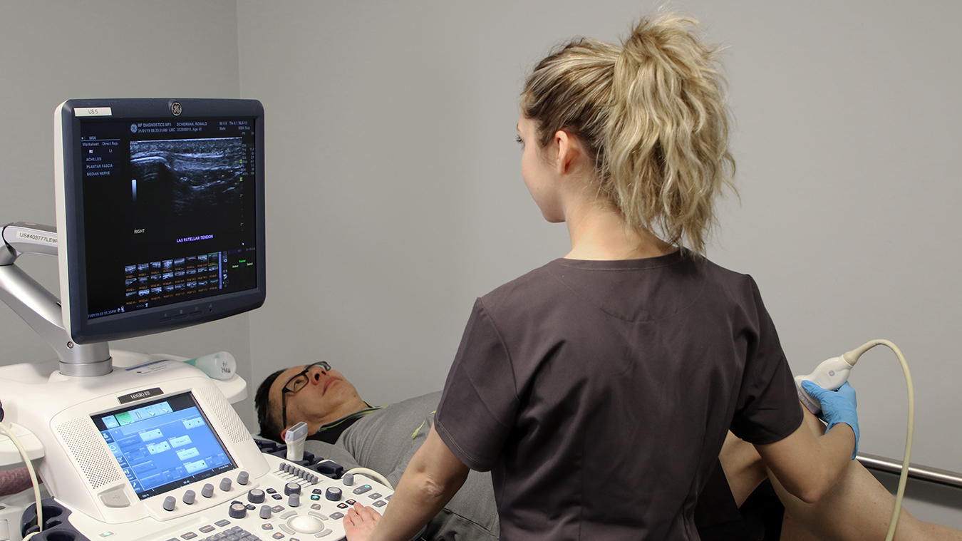
Ultrasound scanners have revolutionized medical imaging, providing doctors with a non-invasive way to view internal organs and tissues in real-time. These devices use high-frequency sound waves to produce images of the body's internal structures, allowing doctors to diagnose and monitor a wide range of medical conditions.
The history of ultrasound dates back to the early 19th century, when scientists first discovered that sound waves could be used to produce images of the body. In the years that followed, researchers worked to refine this technology, developing increasingly sophisticated ultrasound scanners that could produce high-quality images of internal organs and tissues.
One of the key advantages of ultrasound scanners is that they are non-invasive, meaning that they do not require any incisions or injections. This makes them an ideal tool for diagnosing and monitoring a wide range of medical conditions, from heart disease to cancer.
Ultrasound scanners work by emitting high-frequency sound waves into the body, which bounce off internal structures and are then detected by the scanner. These sound waves are then converted into images that can be viewed on a computer screen or printed out for further analysis.
One of the most common uses of ultrasound scanners is in obstetrics, where they are used to monitor the development of fetuses during pregnancy. By using ultrasound, doctors can check for any abnormalities in the fetus, such as structural defects or growth problems.
Ultrasound scanners are also commonly used in cardiology, where they are used to diagnose and monitor heart disease. By using ultrasound, doctors can visualize the heart and its blood vessels, allowing them to detect any blockages or abnormalities that may be present.
In addition to these medical applications, ultrasound scanners are also used in a wide range of other fields, including veterinary medicine, industrial testing, and even in the food industry. In veterinary medicine, ultrasound is often used to diagnose and monitor conditions in animals, while in industrial testing, it is used to detect flaws and defects in materials such as metal and plastic.
Despite their many advantages, ultrasound scanners are not without their limitations. One of the biggest limitations of ultrasound is that it is not always able to produce high-quality images of certain structures, such as bones and air-filled organs. In addition, ultrasound scanners are often unable to penetrate deep into the body, which can make it difficult to visualize certain structures that are located deep within the body.
Despite these limitations, ultrasound scanners have revolutionized medical imaging, providing doctors with a non-invasive way to view internal organs and tissues in real-time. As technology continues to advance, it is likely that ultrasound scanners will become even more sophisticated, allowing doctors to diagnose and monitor a wider range of medical conditions with even greater accuracy and precision.
One of the key advantages of ultrasound scanners is that they are relatively inexpensive compared to other imaging technologies, such as MRI and CT scans. This makes them an ideal tool for use in developing countries and other areas where medical resources may be limited.
Another advantage of ultrasound scanners is that they do not expose patients to ionizing radiation, which can be harmful in large doses. This makes them a safer alternative to other imaging technologies, particularly for pregnant women and children.
Ultrasound scanners are also highly portable, making them an ideal tool for use in remote or rural areas where access to medical facilities may be limited. Portable ultrasound scanners can be easily transported to these areas, allowing doctors to provide medical care to patients who might otherwise have difficulty accessing it.
One of the most exciting developments in ultrasound technology in recent years has been the advent of 3D and 4D ultrasound imaging. These technologies allow doctors to produce three-dimensional images of internal structures, providing a more detailed and accurate view of the body than traditional 2D ultrasound imaging.
In addition to 3D and 4D imaging, researchers are also exploring the use of ultrasound for therapeutic purposes. High-intensity focused ultrasound (HIFU) is a promising new technology that uses high-frequency sound waves to destroy cancerous tumors without the need for surgery or radiation therapy.
Overall, ultrasound scanners have revolutionized medical imaging, providing doctors with a non-invasive, safe, and cost-effective way to view internal organs and tissues in real-time. As technology continues to advance, it is likely that ultrasound scanners will become even more sophisticated, allowing doctors to diagnose and monitor a wider range of medical conditions with even greater accuracy and precision.
In recent years, there has been a growing interest in the use of ultrasound in point-of-care settings, such as emergency departments and intensive care units. Portable ultrasound scanners can be used to quickly diagnose and monitor a wide range of medical conditions, allowing doctors to provide more timely and effective care to their patients.
In addition to its diagnostic uses, ultrasound is also being explored as a tool for guiding medical procedures, such as biopsies and injections. By using ultrasound to guide these procedures, doctors can increase their accuracy and reduce the risk of complications.
Despite its many advantages, ultrasound is not without its limitations. One of the biggest challenges facing ultrasound technology is the need for skilled operators who are able to interpret the images produced by the scanner. In addition, ultrasound scanners are often unable to produce high-quality images in patients who are obese or have a lot of gas in their intestines.
To address these challenges, researchers are working to develop new technologies that can improve the accuracy and reliability of ultrasound imaging. One promising area of research is the use of artificial intelligence (AI) to help interpret ultrasound images. By using machine learning algorithms, AI systems can analyze large amounts of ultrasound data and identify patterns that may be difficult for human operators to detect.
Another area of research is the development of new ultrasound contrast agents that can improve the quality of ultrasound images. These agents work by enhancing the contrast between different tissues in the body, making it easier to visualize internal structures.
In conclusion, ultrasound scanners have revolutionized medical imaging, providing doctors with a non-invasive, safe, and cost-effective way to view internal organs and tissues in real-time. As technology continues to advance, it is likely that ultrasound scanners will become even more sophisticated, allowing doctors to diagnose and monitor a wider range of medical conditions with even greater accuracy and precision. With ongoing research and development, ultrasound technology has the potential to continue improving healthcare outcomes and saving lives around the world.